Prostate Ultrasound
for
Safer Superior Results
when diagnosing the prostate gland

"Prostate ultrasound imaging
has proven far superior to x-ray and other radiation
for helping physicians diagnose the prostate gland.
It has no known side effects.
And, the results are superior."
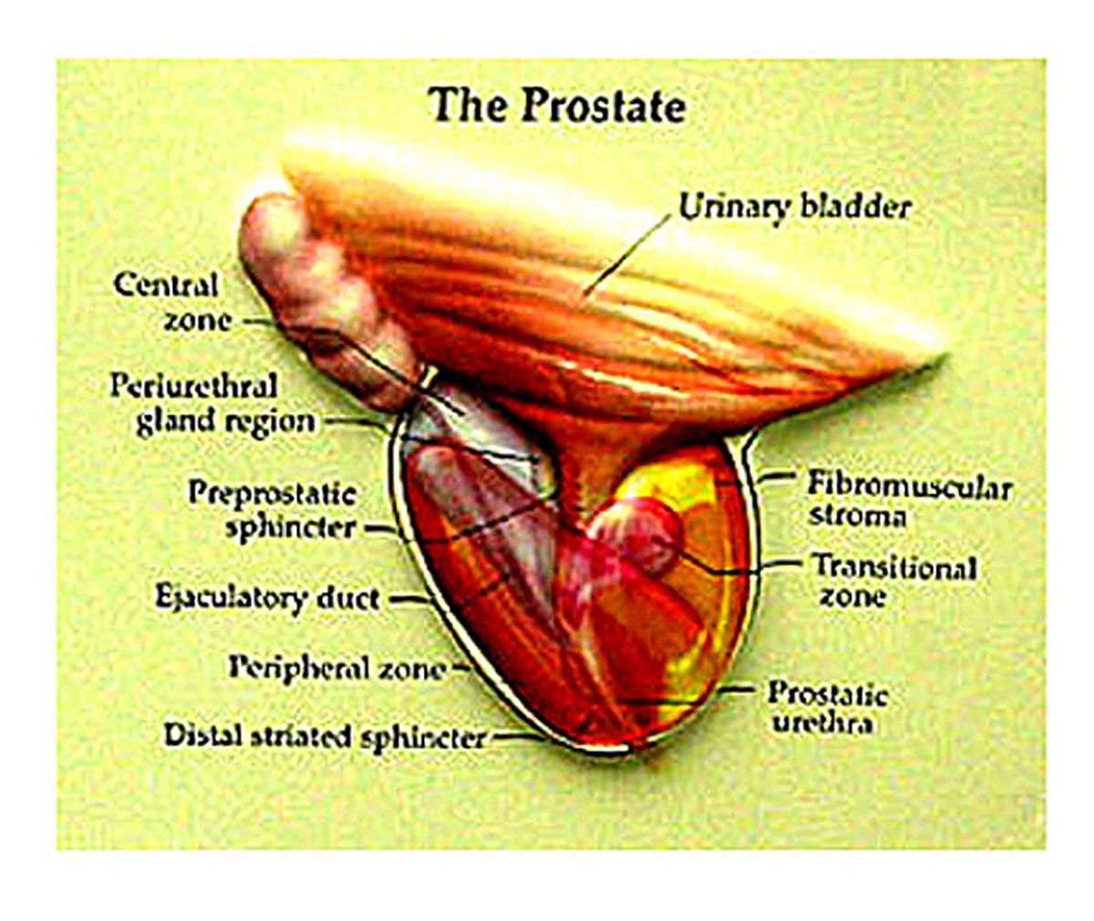
What Is Prostate Ultrasound
and
How Is It Done?
This is a minimally invasive procedure that is done trans rectally (through the rectum). It uses high-frequency sound waves to produce a picture of your prostate gland.
It does not use x-ray or other radiation. And, unlike radiation, standard diagnostic ultrasound has no known side effects. This type of imaging is also in "real time". That means you can see the movement of the prostate gland as well as the blood flowing through the prostate. It is a great aid that helps physicians diagnose the condition of the prostate gland.
Prostate Ultrasound Imaging
is
Safe and Non-Invasive
with
Superior Results
Ultrasound imaging is unlike prostate biopsy (needle biopsy), which is highly invasive and can cause more harm than good. Needle biopsy can release normally contained cancer cells into the blood stream where they can then settle into bones and other organs to cause further cancers.
Ultrasound does not have that associated deadly risk.
"And, unlike x-rays,
sound imaging causes no known health problems
and
can be repeated as often as necessary."
It is also superior to x-ray. It gives a clear picture of soft tissues which do not show up well on x-ray film.
Ultrasound is also much less expensive than an MRI.
What It Can Show You
Transrectal ultrasound imaging of the gland is used to detect abnormalities within it such as:
- Tumors
- Enlargement
- Male infertility
Physicians may want to see an ultrasound image if they feel a lump during a digital rectal exam or if the PSA level was high.
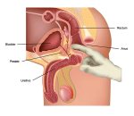
How Is The Procedure Done?
The procedure for ultrasound imaging here is very simple.
Usually you will be given an enema shortly before the test to empty out your bowel. Also you will probably be asked to drink many glasses of water so that your bladder will be full. A full bladder helps to view the prostate.
Then a covered lubricated instrument called a transducer will be inserted into your rectum. Inaudible high frequency sound waves will be emitted from this.
The imaging will usually be performed from different angles to give the best possible view of the gland. The entire exam usually only lasts about 20 minutes.
It Works Just Like The Sonar On A Ship
The way it works is just like sonar on a ship. The sound waves are sent out from the transducer and they bounce back from the objects (organs) they hit. Like an echo. These "echoes" come back like a mirror image, but in sound. The transducer then also records these "echoes".
A computer then translates these sound wave "echoes" into pictures immediately visible on the computer screen.
The pictures are in "real time". That means, with this type of imaging, you can actually see your gland as it is functioning in that moment!
The entire video is usually videotaped so the physician can use it later to diagnose the condition of the gland.
This is the same procedure used in a woman's vagina to view a developing baby.
One
CAUTION
Physicians who use Prostate Ultrasound imaging will typically call for a needle biopsy if any tumors are found. This may or may not be something you wish to do when you know the dangerous and possibly life threatening risks connected to a needle biopsy.
Prostate Ultrasound has many advantages over a needle biopsy when diagnosing tumors.
In any case, Prostate Ultrasound seems to be an effective and
relatively harmless way to get a good look at this gland. And, unlike
x-ray or biopsy, it can safely be used repeatedly.
Be Well.....
~ William
Recent Articles
-
Recent Articles - Page II
Apr 21, 24 08:25 PM
Hello! And, Welcome to page 2 of Recent Articles! The first page was getting really long. So, here is page 2! -
Prostate Massage Success Stories - 2024
Apr 21, 24 08:19 PM
 Prostate massage success stories are becoming as common as blades of grass. The simple, but highly effective techniques
 -
Prostate Massage, The "Nay Sayers"
Apr 21, 24 08:00 PM
 Prostate massage may be all you need to regain excellent prostate health. It's important to avoid the "nay sayers".
 -
The Useless PSA Test and The Potential Dangers - 2024
Apr 04, 24 11:55 PM
 The routine PSA Test is another of the almost totally useless prostate procedures that can unnecessarily scare men.
 -
Natural Prostatitis Pain Relief - 2024
Apr 03, 24 05:22 PM
 Learn the most soothing prostatitis pain treatments that you can use to feel better Right NOW! And, learn how
 -
Fix a Bent Penis Safely and Easily - 2024
Jan 26, 24 06:54 PM
 You can fix a bent penis safely and easily, without surgery, injections, or drugs, 99% of the time. Medically endorsed
 -
Conquering Prostate Problems
Jan 23, 24 06:37 PM
 Conquering prostate problems with correct massage sometimes comes down to just one thing: persistence.
 -
The Prostate Massage Video Series - 2024
Jan 21, 24 06:22 PM
 These Prostate Massage Videos will teach you all you need to know to do the most effective prostate massage possible.
 -
Prostate Massage - Discover the Magical Power - 2024
Jan 19, 24 10:43 PM
 With correct prostate massage you can achieve excellent prostate health again! Whether you're 30 or 90, if you suffer
 -
Prostate Milking - 2024
Jan 19, 24 10:41 PM
 Correct Prostate Milking can bring you great relief if you suffer from prostatitis pain, inflammation, swelling or impo
 -
Correct Prostate Massage Technique - for maximum results - 2024
Jan 13, 24 06:33 PM
 Prostate massage could be the answer to your prayers! Doing it correctly is the difference between success and failure.
 -
So You Have a Bent Penis
Jan 07, 24 08:46 PM
 A bent penis is classified as Peyronies Disease. However, 99% of the time a bent or curved penis is not a disease.


New! Comments
If you'd like to leave me a comment, please use the box below. Thanks! ~ William